Share this
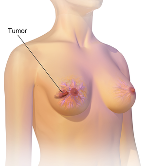
Breast cancer is cancer that develops from breast tissue.[7] Signs of breast cancer may include a lump in the breast, a change in breast shape, dimpling of the skin, milk rejection, fluid coming from the nipple, a newly inverted nipple, or a red or scaly patch of skin.[1] In those with distant spread of the disease, there may be bone pain, swollen lymph nodes, shortness of breath, or yellow skin.[8]
Risk factors for developing breast cancer include obesity, a lack of physical exercise, alcoholism, hormone replacement therapy during menopause, ionizing radiation, an early age at first menstruation, having children late in life or not at all, older age, having a prior history of breast cancer, and a family history of breast cancer.[1][2] About 5–10% of cases are the result of an inherited genetic predisposition,[1] including BRCA mutations among others.[1] Breast cancer most commonly develops in cells from the lining of milk ducts and the lobules that supply these ducts with milk.[1] Cancers developing from the ducts are known as ductal carcinomas, while those developing from lobules are known as lobular carcinomas.[1] There are more than 18 other sub-types of breast cancer.[2] Some, such as ductal carcinoma in situ, develop from pre-invasive lesions.[2] The diagnosis of breast cancer is confirmed by taking a biopsy of the concerning tissue.[1] Once the diagnosis is made, further tests are done to determine if the cancer has spread beyond the breast and which treatments are most likely to be effective.[1]
The balance of benefits versus harms of breast cancer screening is controversial. A 2013 Cochrane review found that it was unclear if mammographic screening does more harm than good, in that a large proportion of women who test positive turn out not to have the disease.[9] A 2009 review for the US Preventive Services Task Force found evidence of benefit in those 40 to 70 years of age,[10] and the organization recommends screening every two years in women 50 to 74 years of age.[11] The medications tamoxifen or raloxifene may be used in an effort to prevent breast cancer in those who are at high risk of developing it.[2] Surgical removal of both breasts is another preventive measure in some high risk women.[2] In those who have been diagnosed with cancer, a number of treatments may be used, including surgery, radiation therapy, chemotherapy, hormonal therapy, and targeted therapy.[1] Types of surgery vary from breast-conserving surgery to mastectomy.[12][13] Breast reconstruction may take place at the time of surgery or at a later date.[13] In those in whom the cancer has spread to other parts of the body, treatments are mostly aimed at improving quality of life and comfort.[13]
Outcomes for breast cancer vary depending on the cancer type, the extent of disease, and the person’s age.[13] The five-year survival rates in England and the United States are between 80 and 90%.[14][4][5] In developing countries, five-year survival rates are lower.[2] Worldwide, breast cancer is the leading type of cancer in women, accounting for 25% of all cases.[15] In 2018, it resulted in 2 million new cases and 627,000 deaths.[16] It is more common in developed countries[2] and is more than 100 times more common in women than in men.[14][17]

The development of breast buds is one of the first signs of puberty in a girl.
The development of breast buds is one of the first signs of puberty in a girl. The beginning of adult breast development is called thelarche. As the breasts start to develop, a girl will have small, firm lumps under the nipples called breast buds, which may sometimes be tender. In some cases, one breast may start to develop weeks or months before the other. The breast tissue continues to get larger and become less firm over the next few years. It is normal for Caucasian girls to begin developing breast buds as young as seven years old and African American girls could be as young as six years old. It is advised to consult a pediatrician to evaluate breast bud development and check for other signs of puberty.
A girl usually attains menarche (gets her first period) around two years after breast development begins. The average age for girls in the United States to start their periods is anywhere between 9 to 15 years. Girls usually have a similar pattern to their mothers when it comes to beginning their period. If the mother started her period early, the daughter is more likely to start early. However, this is not true for everyone. Nutrition, medical illnesses, long-term medication and other environmental factors can also influence the onset of puberty.
What is precocious puberty?
Precocious puberty is when the signs of puberty begin before the age of seven or eight years in girls and before the age of nine years in boys. When this happens, it is advised to consult a pediatrician to rule out any serious causes and help manage the effects of precocious puberty.
The effects of precocious puberty
Early puberty can affect the child emotionally and socially. Children with precocious puberty may be confused or conscious about the changes in their bodies. They may have changes in emotions and behavior, and they need guidance on how to navigate through these changes. Girls could be concerned about getting periods and can become moody and irritable. Boys may become more aggressive and develop a sex drive inappropriate for their age. Girls who attain puberty early may be at a higher risk of sexual abuse, hence it is important to educate children. It may also be a good idea to seek the help of the teacher at school, a counselor or a psychologist.
Precocious puberty causes a growth spurt in the child, making them taller than their peers. However, their bone growth stops at an earlier age than other kids. They stop growing in height sooner and end up shorter in height than they would have otherwise been.
What are the causes of precocious puberty?
The onset of puberty is triggered by the hypothalamus, an area in the brain that sends signals to the pituitary gland (a small gland at the base of the brain) to release hormones that stimulate the ovaries (in girls) or testicles (in boys) to produce sex hormones.
Precocious puberty in girls is often due to the hypothalamus sending these signals earlier than it should. This may run in families and usually not a medical problem.
Rarely, precocious puberty may occur due to a more serious medical problem, such as a brain tumor or trauma. Thyroid or ovarian disorders can also trigger early puberty. Hence, it is advised to consult a pediatrician to rule out anything serious and help manage precocious puberty in the child.
How is precocious puberty treated?
The treatment goals are to stop or reverse sexual development or stop rapid bone growth and maturation. This growth spurt may lead to short stature as an adult. Depending on the cause of precocious puberty, treatment may involve treating the underlying disease and lowering the high levels of sex hormones with medication to stop sexual development.
Hormone treatment with drugs called luteinizing hormone releasing hormone (LHRH) analogs may be used in precocious puberty. LHRH analogs are synthetic hormones that block the body’s production of sex hormones, which cause early puberty. They are typically safe and don’t cause side effects in children. It is also extremely important to address the child’s mental health at this stage. Parents should help them cope with their changes physically and mentally by taking help from family, friends, teachers, a counselor or a psychologist.

Things Young Women Should Know About Breast Cancer
Breast Cancer in Women Under 40

Each year, around 12,000 women under age 40 will be diagnosed with breast cancer, making up less than 5% of all breast cancer cases, and it is the most common cancer found in women in this age group.
Throughout her lifetime, a woman has a 1 in 8 risk of developing breast cancer. No matter what your age you need to be aware of risk factors. In many cases of breast cancer early diagnosis is the key to survival.
This slideshow will tell you 10 things every young woman should know about breast cancer.
What is Breast Cancer?

Breast cancer is the most common cancer in American women, and it is the second most common cause of cancer deaths in women. (Lung cancer still kills almost 4 times as many women each year as breast cancer.) Breast cancer occurs rarely in men as well. There are about 230,000 new cases of breast cancer diagnosed in women the U.S. each year, and about 2,300 new cases diagnosed in men.
To understand breast cancer, it’s important to learn the anatomy of the breast. Most of the breast is comprised of fatty (adipose) tissue, and within that are ligaments, connective tissue, lymph vessels and nodes, and blood vessels. In a female breast there are 12-20 sections within it called lobes, each made up of smaller lobules that produce milk. The lobes and lobules are connected by ducts, which carry the milk to the nipple.
The most common type of breast cancer is cancer of the ducts, called ductal carcinoma, that accounts for just over 80% of all breast cancers. Cancer of the lobes (lobular carcinoma) makes up just over 10% of cases. The rest of the breast cancers have characteristics of both ductal and lobular carcinomas, or have unknown origins.
1. Know Your Breasts

While women under 40 only make up less than 5% of diagnosed breast cancer cases, breast cancer is a leading cause of death among young women age 15-34. It is important to know your breasts. Know how they feel, and have your doctor teach you how to do a proper breast self-exam, if you choose, to help you notice when there are changes that need to be examined by a doctor.
2. Know The Risk Factors

Younger women may have a higher risk for developing breast cancer with the following risk factors:
- Certain inherited genetic mutations for breast cancer (BRCA1 and/or BRCA2)
- A personal history of breast cancer before age 40
- Two or more first-degree relatives (mother, sister, daughter) with breast cancer diagnosed at an early age
- High-dose radiation to the chest
- Early onset of menstrual periods (before age 12)
- First full-term pregnancy when you are over 30 years old
- Dense breasts
- Heavy alcohol consumption
- Obesity
- Sedentary lifestyle
- High intake of red meat and poor diet
- Race (Caucasian women have a higher risk)
- Personal history of endometrium, ovary, or colon cancer
- Recent oral contraceptive use
3. Breast Changes to Watch

Watch for changes to your breasts, and if you notice any of the following, see your doctor:
- A lump in or near your breast or under your arm
- Changes in the size or shape of your breast
- Dimpling, puckering, or bulging of the skin
- A nipple that has changed position or an inverted nipple (pushed inward instead of sticking out)
- Skin redness, soreness, rash
- Swelling
- Nipple discharge (could be a watery, milky, or yellow fluid, or blood)
Normal breast tissue may be lumpy, which is why it is important to know how your breasts normally feel. Most lumps are not cancer. Many women choose to perform breast self-exams so they will know if a new lump appears or an existing lump changes size. However, breast self-exams are not a substitute for mammograms.
These changes may not necessarily indicate that you have breast cancer, but they could and should be evaluated.
4. Be Persistent and Speak Up

Be your own health advocate and make sure you mention any breast changes or lumps to your doctor. Some patient concerns are dismissed because they are “too young” to have breast cancer. If you think you feel something, seek answers. Don’t be afraid to get a second opinion and more information.
5. Find The Right Doctor

If you are diagnosed with breast cancer, it’s important to find the right medical team to work with you. It may be tempting to stick with your first doctor, but it’s always a good idea to get a second opinion and make sure you are seeing the right specialists for your type of cancer. You may see several different types of oncologists (cancer specialists), including medical, surgical, and radiation oncologists. The medical specialists you see should be well versed on all the new treatments and approaches including genetics and neoadjuvant therapy (chemotherapy before surgery). Make sure your doctors know the National Comprehensive Cancer Network (NCCN) treatment guidelines which determine treatment based on stage of the disease and prognostic factors of the tumor that are considered the gold standard. You may also want a care-manager or caseworker to help you on your journey.
6. Know Your Medical History

It is important to know your family history and share it with your doctor. Women with a first-degree relative (mother, sister, daughter) with breast cancer have nearly twice the risk of being diagnosed with breast cancer as a woman who has no family history. Tell your doctor which family member(s) had breast cancer or other breast diseases, and how old they were when diagnosed.
7. Seek a Second Opinion

Most doctors will suggest getting a second opinion, and even if they do not, it is always a good idea. Most insurance will cover it. It’s important to seek a specialist in breast cancer who is up to date on the latest treatments and can help you make the best decisions on how to proceed. You may discuss your diagnosis with another pathologist who can review your breast tissue slides and confirm a diagnosis, or another medical oncologist, surgical oncologist, or radiation oncologist to determine the best treatment choices.
8. Know It’s OK to Ask Questions

Ask questions! You should be an active participant in your care. Your medical team should explain to you any medical terms you do not understand, explain your treatment choices, possible side effects, and expected outcome. Ask for references to additional specialists you can talk to so you can learn more about your breast cancer. If you have not yet been diagnosed with breast cancer but are at high risk, ask your doctors about testing and any preventive measures you can take.
Also don’t be afraid to ask family and friends for support. Seek support groups with other people who are going through what you are, or who have gone through it. Bring a close friend or family member to your appointments to both take notes, or record your visit, and to encourage you to request clarification if anything is unclear. Express your feelings and concerns.
9. Do Some Research

If you are diagnosed with breast cancer, learn about your specific diagnosis. Understand what terms such as stage and grade mean, and how they impact your treatment options.
10. Network With Other Young Women

It can feel isolating to be diagnosed with breast cancer at a younger age, but there is support available and it can be helpful to connect with other women your age who are going through what you are, or who have beat breast cancer. You can start by asking your doctor about any local support groups. In addition, you can find support groups by searching online.
Breast Cancer Prevention for Young Women

If you are a young woman there are some risk factors for breast cancer you can avoid.
- Don’t smoke
- Exercise regularly
- Eat a healthy diet, with an emphasis on plant foods
- Limit consumption of red meats and processed meats
- Maintain a healthy weight
- Limit or avoid alcohol consumption
- If possible, avoid shift work, especially at night
Changing your lifestyle and habits may not completely prevent you from getting cancer but it can lower your risk, especially if you have some unavoidable risk factors already such as a genetic history.
Additional Information on Breast Cancer
For more information about Breast Cancer, please consider the following:
- American Cancer Society
- National Breast Cancer Foundation, Inc.
- BreastCancer.org
- Susan G. Komen
- National Cancer Institute
Health SolutionsFrom Our Sponsors
- Penis Curved When Erect
- Could I have CAD?
- Treat Bent Fingers
- Treat HR+, HER2- MBC
- Tired of Dandruff?
- Life with Cancer
Breast Cancer: Where It Can Spread
When Cancer Goes Beyond Your Breast

If your doctor told you that your breast cancer has spread to other parts of your body, it’s at a more advanced stage than if it’s only in your breasts. How far it has spread is one of the things your doctor will consider when they tell you the “stage” of your cancer. It’s considered “metastatic” if it has spread far from your breasts. Every case is different. For some women, it becomes something they live with for a long time. For others, focusing on pain management and quality of life is the main goal.
Most Common Places It Spreads

It’s still breast cancer, even if it’s in another organ. For example, if breast cancer spreads to your lungs, that doesn’t mean you have lung cancer. Although it can spread to any part of your body, there are certain places it’s most likely to go to, including the lymph nodes, bones, liver, lungs, and brain.
Lymph Nodes

The lymph nodes under your arm, inside your breast, and near your collarbone are among the first places breast cancer spreads. It’s “metastatic” if it spreads beyond these small glands to other parts of your body. When you’re diagnosed with breast cancer, your doctor should check lymph nodes near the tumor to see if they’re affected. The lymph system helps drain bacteria and other harmful things from your body. You might not notice symptoms if your breast cancer is in these nodes.
Bones

When breast cancer is in your bones, pain is usually the first symptom. It can affect any bone, including the spine, arms, and legs. Sometimes the bone may be weak enough to break, but treatment often prevents that. If the cancer involves your spine, it can also cause problems with incontinence or going to the bathroom. You might also have numbness or weakness in a part of your body, like an arm or leg. That happens when there’s pressure on the nerves of the spinal cord.
Liver

If breast cancer spreads to your liver, you may have pain in your belly that doesn’t go away, or you might feel bloated or full. You might also lose your appetite and lose weight. You may notice that your skin and the whites of your eyes are turning yellow, which is called jaundice. That happens because your liver isn’t working right.
Lungs

Breast cancer can spread to the lungs or to the space between the lung and the chest wall, making fluid build up around the lung. Symptoms can include shortness of breath, a cough that won’t go away, and chest pain. Some people lose their appetite, leading to weight loss.
Brain

It’s possible for breast cancer to spread to the brain. That can cause headaches that throw off your balance and make falls more likely. You may have numbness or weakness in one part of your body. You might act differently, or you could feel confused or have seizures.
Treatments

You may need surgery, chemotherapy, radiation, and medications. The drugs your doctor recommends will depend on your type of breast cancer. For instance, if your breast cancer is HER2 positive, in which a certain protein drives the growth, your doctor may choose targeted therapy as part of your treatment. Pain management is also key so you can feel as well as possible.
Health SolutionsFrom Our Sponsors
- Penis Curved When Erect
- Could I have CAD?
- Treat Bent Fingers
- Treat HR+, HER2- MBC
- Tired of Dandruff?
- Life with Cancer
Breast Cancer Help During After Treatment
Acupuncture

In this ancient practice, very thin needles are put on points of your body. Research shows it boosts your immune system and releases natural painkillers. It may also curb side effects of cancer treatment like nausea, pain, fatigue, and anxiety.
But don’t skip doctor visits because you’re getting acupuncture. In fact, make sure your doctor knows you’re thinking about it before you try it. There can be side effects and in some cases, acupuncture isn’t recommended.
Massage

Some hospitals have massage therapists on staff. Massage can lessen pain. It can also help you relax before a lumpectomy, mastectomy, or breast reconstruction. After surgery, a specialized massage given by a specially trained therapist may help lessen swelling. Talk with your doctor about it. They may mention “lymph drainage techniques.” If so, that’s what they’re talking about.
Yoga

This form of exercise helps connect breath to movement. It slows your heart rate, blood pressure, and brain waves. Women who take yoga classes while having radiation for breast cancer say they feel less tired and stressed. If you can do it regularly, yoga may also lessen inflammation. Make sure to share your medical history with your instructor so they’ll know best how to help you.
Tai Chi

This age-old Chinese martial art combines slow, graceful body movements with breathing and meditation. Relaxing the mind this way may help strengthen the body. Tai chi may lessen inflammation in people with breast cancer. Women who practice tai chi for an hour, 3 three times a week also feel better about their health and their lives.
Art Therapy

You don’t need to be a good artist to reap the benefits of art therapy. When you draw, paint, sculpt or craft, it gives you a chance to express fears and other feelings you may not want to talk about. Working with a trained art therapist can help you feel better about your treatment. It’s also been shown to ease anxiety and depression. It can also help your self-esteem.
Makeovers

Side effects like hair loss and skin problems hit many women hard. Some people in treatment don’t like to leave their house because they don’t like how they look. When you feel better about your appearance, it can improve your quality of life and help you stay strong. Find a program in your area that will fit you for a wig, give you a makeover, or give you a bra for your new shape. Some are free.
Equine-Assisted Therapy

Horses are natural therapists. And since they mirror the body language of people around them, horses can help you become more aware of your feelings. Learning to care for and ride a horse builds confidence. In one small study, people who participated in therapy with horses felt less stress. We’re not really sure why, but it could be worth a try.
Music Therapy

Listening to your favorite tunes has probably helped you get over a breakup or power through a workout. This ability to connect you to your feelings is why music can also help during treatment. Studies show a program with a trained music therapist can drop pain levels, improve state of mind, and reduce worry for people with breast cancer. Ask your doctor to recommend one.
Write About It

If you write down your feelings about breast cancer, from your hopes to your biggest fears, you may notice fewer physical symptoms. Writing in a journal can also boost your mood and help you see the progress you’re making. Don’t worry about spelling or handwriting. Just let go and focus on your thoughts and goals.
Support Groups

No matter how much care you get from friends and family, a support group may also help. Time with other women who are going through the same thing can help you feel less alone. You may talk more about your problems since other group members know what you’re dealing with. You can also ask for advice, like what to expect during different stages of treatment or how to handle side effects.
Health SolutionsFrom Our Sponsors
- Penis Curved When Erect
- Could I have CAD?
- Treat Bent Fingers
- Treat HR+, HER2- MBC
- Tired of Dandruff?
- Life with Cancer
Breast Cancer Awareness: Symptoms and Treatment
Breast Cancer Awareness

The outlook for women with breast cancer is improving constantly. Due to increased awareness, opportunities for early detection, and treatment advances, survival rates continue to climb. In the U.S., October is Breast Cancer Awareness Month, and the campaign is designed to increase breast cancer awareness. There are many organizations that support Breast Cancer Awareness Month and provide assistance within early detection plans. Organizations also put together breast cancer fundraisers such as walks and events that support breast cancer research and help fund patients with socio-economic disadvantages.
Breast Cancer Symptoms

Breast cancer may or may not cause symptoms. Some women may discover the problem themselves, while others may have the abnormality first detected on a screening exam. Common breast cancer symptoms, when they occur, include the following:
- Non-painful lumps or masses
- Lumps or swelling under the arms
- Nipple skin changes or discharge
- Noticeable flattening or indentation of the breast
- Change in the nipple
- Unusual discharge from the nipple
- Changes in the feel, size, or shape of the breast tissue
Types of Breast Cancer

Inflammatory Breast Cancer
Inflammatory breast cancer is a rare type of cancer that often does not cause a breast lump or mass. As seen in this photo, it often causes thickening and pitting of the skin, like an orange peel. The affected breast may also be larger or firmer, tender, or itchy. A skin rash or reddening of the skin is common. These changes are caused by cancer cells blocking lymph vessels in the skin. Inflammatory cancer of the breast typically has a fast growth rate.
Invasive Ductal Carcinoma
Invasive (or infiltrating) ductal carcinoma (IDC) is the most common type of breast cancer. About 80% of all breast cancers are invasive ductal carcinomas. Invasive ductal carcinoma refers to cancer that has broken through the wall of the milk ducts and has invaded the breast tissues. Invasive ductal carcinoma can spread to the lymph nodes and possibly to other areas of the body.
Ductal Carcinoma in Situ (DCIS)
Ductal carcinoma in situ (DCIS) is considered to be a non-invasive or pre-invasive breast cancer. Ductal means that the cancer starts inside the milk ducts, carcinoma refers to any cancer that starts in the skin or other tissue (including breast tissue) that line or cover the internal organs, and in situ means “in its original place.” The difference between DCIS and invasive cancer is that in DCIS, the cells have not spread through the walls of the milk ducts into the surrounding breast tissue. DCIS is considered a ‘pre-cancer’, but some cases can transform into more invasive cancers.
Invasive Lobular Carcinoma
Invasive (or infiltrating) lobular carcinoma (ILC) is the second most common type of breast cancer after invasive ductal carcinoma. Lobular means that the cancer started in the milk-producing lobules, which empty out into the ducts that carry milk to the nipple. Invasive lobular carcinoma refers to cancer that has broken through the wall of the lobule and begun to invade the breast tissues. Invasive lobular carcinoma can spread to the lymph nodes and possibly to other areas of the body.
Mucinous Carcinoma
Mucinous (or colloid) carcinoma of the breast is a rare form of invasive ductal carcinoma. In this type of cancer, the tumor is composed of abnormal cells that “float” in pools of mucin, part of the slimy, slippery substance known as mucus. Mucus lines most of the inner surface of our bodies, such as our digestive tract, lungs, liver, and other vital organs. Breast cancer cells can produce some mucus. In mucinous carcinoma, mucin becomes part of the tumor and surrounds the breast cancer cells.
“Pure” mucinous carcinomas make up only 2-3% of invasive breast cancers. Approximately 5% of invasive breast cancer tumors have a mix of mucinous components in addition to other types of breast cancer cells.
Triple-Negative Breast Cancers
Testing negative for estrogen receptors (ER-), progesterone receptors (PR-), and HER2 (HER2-) on a pathology report means the cancer is “triple-negative”. These negative results indicate the growth of the cancer is not supported by the hormones estrogen and progesterone, nor by the presence of too many HER2 receptors. Therefore, triple-negative breast cancer does not respond to hormonal therapy (such as tamoxifen or aromatase inhibitors) or therapies that target HER2 receptors, such as Herceptin. However, other medicines can be used to treat triple-negative breast cancer.
Paget’s Disease of the Nipple
Paget’s disease of the nipple is a rare form of breast cancer in which cancer cells collect in or around the nipple. The cancer usually affects the ducts of the nipple first then spreads to the nipple surface and the areola. A scaly, red, itchy, and irritated nipple and areola are signs of Paget’s disease of the nipple. One theory for the cause of Paget’s disease is that the cancer cells start growing inside the milk ducts within the breast and then break through to the nipple surface. Another possibility is that the cells of the nipple itself become cancerous.
Causes of Breast Cancer

Certain genes control the life cycle—the growth, function, division, and death—of a cell. When these genes are damaged, the balance between normal cell growth and death is lost. Normal breast cells become cancerous because of changes in DNA structure. Breast cancer is caused by cellular DNA damage that leads to out-of-control cell growth.
Causes of Breast Cancer: Genetics & Mutations
Inherited genes can increase the likelihood of breast cancer. For example, mutations of genes BRCA1 and BRCA2 (linked to an increased risk of breast and ovarian cancers) can inhibit the body’s ability to safeguard and repair DNA. Copies of these mutated genes can be passed on genetically to future generations, leading to a genetically-inherited increased risk of cancer.
Causes of Breast Cancer: Lifestyle
Lifestyle choices can lead to breast cancer as well. Eating a poor diet, inactivity, obesity, heavy alcohol use, tobacco use including smoking, and exposure to chemicals and toxins are all associated with a greater breast cancer risk.
Causes of Breast Cancer: Medical Treatment
Medical treatment with chemotherapy, radiation, or immunosuppressive drugs used to decrease the spread of cancer throughout the body can also cause damage to healthy cells. Some “second cancers,” completely separate from the initial cancer, have been known to occur following aggressive cancer treatments. Radiation theraspy to the chest to treat other conditions or cancers also increases the risk of developing breast cancer.
Mammograms and Breast Cancer Prevention

Early detection of breast cancer is the key to survival. Mammograms are X-rays of the breast that can detect tumors at a very early stage, before they would be felt or noticed otherwise. During a mammogram, your breasts are compressed between two firm surfaces to spread out the breast tissue. Then an X-ray captures black-and-white images of your breasts that are displayed on a computer screen and examined by a doctor who looks for signs of cancer. 3D mammograms, or breast tomosynthesis, is a breast imaging procedure that also uses X-rays to produce images of breast tissue in order to detect abnormalities.
Breast Cancer Prevention: Breast MRI and Ultrasound

Breast MRI
MRI (magnetic resonance imaging) is a technology that uses magnets and radio waves to create detailed, 3D images of the breast tissue. Before the test you may be injected with a contrast solution (dye) through an intravenous line in the arm. The contrast solution will allow potential cancerous breast tissue to show more clearly. Radiologists are able to see areas that could be cancerous because the contrast tends to be more concentrated in areas of cancer growth.
During a breast MRI the breasts are exposed as the patient lies flat on a padded platform with cushioned openings for the breasts. A breast coil surrounds each opening and works with the MRI unit to create the images. MRI imaging is a painless diagnostic tool. The test takes between 30 and 45 minutes.
Ultrasound
Sometimes a breast ultrasound is ordered in addition to a mammogram. An ultrasound can demonstrate fluid-filled cysts that are not cancerous. Ultrasounds may also be recommended for routine screening tests in some women at a higher risk of developing breast cancer. During a breast ultrasound a small amount of water-soluble gel is applied to the skin over the area to be examined. Then, a probe is gently applied against the skin. You may be asked to hold your breath, briefly several times. The breast ultrasound takes about 10 minutes to complete.
Breast Cancer Prevention: Breast Self-Exams

Experts recommend that women be aware of their breasts and notice any changes, rather than performing checks on a regular schedule. Women who choose to do self-exams should be sure to discuss the technique with their doctor.
What is a Breast Self-Exam?
A breast self-exam is a way to check your breasts for changes such as lumps or thickenings. Early breast cancer detection can improve your chances of surviving the disease. Any unusual changes discovered during the breast self-exam should be reported to your doctor.
Lump in Breast: Could it be Cancer?

Remember that the majority (about 80%) of breast lumps are not due to cancer. Cysts, benign tumors, or changes in consistency due to the menstrual cycle can all cause benign breast lumps. Still, it’s important to let your doctor know about any lumps or changes in your breast that you find.
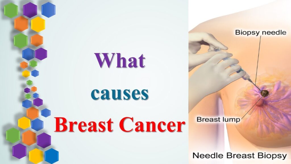
Breast Cancer Biopsy

A biopsy is the most certain way to determine whether a breast lump is cancerous. Biopsies may be taken through a needle or through a minor surgical procedure. The results can also determine the type of breast cancer that is present in many cases (there are several different types of breast cancers). Treatments are tailored to the specific type of breast cancer that is present.
Needle Biopsies
A needle biopsy uses a hollow needle to remove tissue or cell samples from the breast. A pathologist studies the samples under a microscope to see if they contain cancer. There are two types of needle biopsies: core need biopsy and fine needle aspiration (fine needle biopsy).
Core Needle Biopsy
If a lump can be felt in the breast (palpable mass), a core needle biopsy may be performed. The doctor will use a small amount of local anesthetic to numb the skin and the breast tissue around the area. The doctor will insert the needle and remove a small amount of tissue to be examined.
Ultrasound-Guided Core Needle Biopsy
This is one type of biopsy for lumps or abnormalities that cannot be felt (nonpalpable mass). A core needle is placed into the breast tissue and ultrasound helps confirm the exact location of the potential cancer so the needle is placed correctly. Tissue samples are then taken through the needle. Ultrasound can see the difference between cysts and solid lesions.
MRI-Guided Core Needle Biopsy
For this test, you will be given a contrast agent through an IV. Your breast will be numbed and compressed and several MRI images will be taken. The MRI images will guide the doctor to the suspicious area. A needle will be used in the biopsy device to remove tissue samples with a vacuum assisted probe.
Stereotactic Biopsy
If the lump is nonpalpable you could be also given a stereotactic biopsy. Using a local anesthetic, the radiologist makes a small opening in the skin. A needle is placed into the breast tissue, and imaging studies help confirm the exact placement. Tissue samples are taken through the needle.
Surgical Biopsies
A surgeon makes a cut (incision) in the breast to remove tissue.
Open Excisional Biopsy
This surgery removes an entire lump and the issue is examined under a microscope. If a section of normal breast tissue is taken all the way around a lump, it is called a lumpectomy. In this procedure, a wire is put through a needle into the area to be biopsied. The X-ray helps to make sure it is in the right location and a small hook at the end of the wire keeps it in position. The surgeon uses the wire as a guide to locate the suspicious tissue.
Incisional Biopsy
An incisional biopsy is very similar to an excisional biopsy, but less tissue is removed. Local anesthetic will be used, and you will also get IV sedation. An incisional biopsy removes part of the tumor, which means that more surgery may be needed to remove the remaining cancer.
Biopsy Results: Hormone-Sensitive Breast Cancer

A biopsy can tell whether the breast cancer has receptors for estrogen (ER-positive) and/or progesterone (PR-positive), indicating which hormone stimulates tumor growth. About two-thirds of breast cancers are hormone receptor-positive. Medications can be given that act to help prevent growth of the tumor from stimulation by these hormones.
ER-positive breast cancer is sensitive to estrogen, whereas PR-positive breast cancer is sensitive to progesterone. Both ER-positive and PR-positive breast cancers may respond to hormone therapy. Hormone receptor (HR) negative is a type of cancer that does not have hormone receptors and will not be affected by hormone blocking treatments.
Biopsy Results: HER2-Positive Breast Cancer

HER-2 (human epidermal growth factor receptor 2) is a protein that is expressed at a high level by about 20% of breast cancers. Having this receptor means the cancer tends to grow and spread faster than other forms of breast cancer. However, there are special targeted treatments available for this type of tumor.
Treatments specifically for HER2-positve breast cancer include:
- Herceptin (trastuzumab)
- Kadcyla (ado-trastuzumab emtansine)
- Perjeta (pertuzumab)
- Tykerb (lapatinib)
Breast Cancer Stages

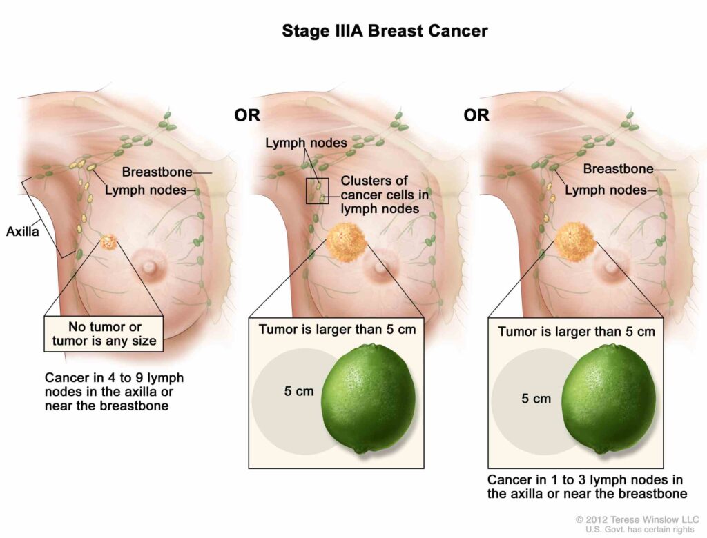
Breast cancer stages are classified according to cancer tumor size, location, and extent of spread. Staging helps doctors determine the prognosis and treatment for cancer. The TNM staging system classifies breast cancers according to the following criteria:
- Tumor (T): Primary tumor size and/or extent
- Nodes (N): Spread of cancer to lymph nodes in the regional area of the primary tumor
- Metastasis (M): Spread of cancer to distant sites away from the primary tumor
Breast Cancer Survival Rates

Breast cancer survival depends upon a number of factors. Cancers that are found early are often localized to the breast. Statistics on the survival rate of breast cancer are often given as 5-year survival rates. The 5-year survival rate is the percentage of people who live at least 5 years after being diagnosed with breast cancer. According to the American Cancer Society, women with early stage (stage 1) breast cancer have a 5-year survival rate of 100%. Women with breast cancer that has spread to distant sites in the body (stage 4) have only a 22% chance of surviving 5 years; but this rate can improve as treatment advances are made.
Breast Cancer Treatments: Surgery

Breast-conserving surgery removes the cancer and some healthy tissue around it, but not the breast. Some lymph nodes under the arms may be removed for biopsy. If the cancer is near the chest wall, part of it may be removed. Breast-conserving surgery is also known as breast-sparing surgery, lumpectomy, partial mastectomy, quadrantectomy, and segmental mastectomy.
Mastectomy
Mastectomy is the removal of the entire breast and all the surrounding tissue and possibly nearby tissues. There are different mastectomy surgeries available, depending on how much additional tissue is removed.
Breast Cancer Treatments: Radiation Therapy

High-energy beams of localized radiation are used to kill targeted cancer cells. Radiation therapy can be used after breast cancer surgery, or it may be used in addition to chemotherapy for widespread cancer. This treatment does have side effects, which can include swelling of the area, tiredness, or a sunburn-like effect. There are two ways to administer radiation therapy.
External Beam Radiation
A beam of radiation is focused onto the affected area by an external machine. The treatment is usually given five days a week for five to six weeks.
Brachytherapy
This form of radiation involves radioactive seeds or pellets that are implanted into the breast next to the cancer.
Breast Cancer Treatments: Chemotherapy

Chemotherapy drugs are given to kill cancer cells that are located anywhere in the body. It can be administered by a slow IV infusion, by pill, or by a brief IV injection, depending upon the drug. Sometimes chemotherapy is given after surgery to help prevent the cancer from recurring (adjuvant therapy). Side effects of chemotherapy can include an increased risk of infection, nausea, fatigue, and hair loss.
Adjuvant Chemotherapy
If all visible cancer has been removed, there is still the possibility that cancer cells have broken off or are left behind. Adjunct chemotherapy is given to assure that these small amounts of cells are killed. Since some women have a very low risk of recurrence even without chemotherapy, it is not given in all cases.
Neoadjuvant Chemotherapy
Neoadjuvant chemotherapy is given before surgery. There is no correlation between neoadjuvant chemotherapy and long-term survival, but there are advantages to see if the cancer responds to the chemotherapy before surgical removal. This can also reduce the size of the cancer and allow for a less extensive surgery in some patients.
Chemotherapy for Advanced Breast Cancer
Chemotherapy can be used if the cancer has metastasized to distant sites in the body. In this case, doctors will determine the most appropriate treatment.
Chemotherapy Side Effects
Different drugs cause different side effects. Certain types of chemotherapy have specific side effects, but each patient’s experience is different. The following are common side effects of chemotherapy:
- Fatigue
- Pain (headaches, muscle pain, stomach pain, and pain from nerve damage)
- Mouth and throat sores
- Diarrhea
- Nausea and vomiting
- Constipation
- Blood disorders
- Changes in thinking and memory
- Sexual and reproductive issues
- Appetite loss
- Hair loss
- Permanent damage to the heart, lung, liver, kidneys, or reproductive system

Some breast cancer cells are activated by female hormones estrogen and/or progesterone (ER- and PR-positive breast cancers). Hormone therapy can stop or slow the growth of hormone receptor-positive tumors by blocking the cancer cells from receiving the hormones they need to grow. Hormone therapy is usually given after surgery, but it can also be given to reduce the chance of developing breast cancer in women at high risk.
Targeted Drug Therapies for Breast Cancer

Targeted therapies are newer treatments for breast cancer patients. They utilize specific proteins within cancer cells, like the HER-2 protein. Targeted therapies can stop the HER-2 protein from stimulating tumor growth in cancer cells that have this protein. Targeted therapies have fewer side effects than traditional chemotherapy because they only target cancer cells. They are often used in combination with chemotherapy.

Breast cancer treatment can be both physically and emotionally exhausting. There are many changes taking place that may be difficult to cope with. “Chemobrain” is a term coined to describe the mental changes caused by chemotherapy treatment. Patients have experienced memory deficits and the inability to focus. Breast cancer treatments can also leave patients fatigued, which is normal.
It can be difficult to keep up with activities of daily life, and make patients feel isolated or overwhelmed. Friends and family can be invaluable sources of support and assistance during this time. Some people choose to join a local or an online support group to share their experiences and spread breast cancer awareness.
Breast Reconstructive Surgery

Many women opt to have reconstructive surgery after breast cancer surgery. Reconstructive procedures use implants or tissues obtained from other locations in the body. These procedures can be done at the time of mastectomy, or they may be performed months or even years later.
Implants
A tissue expander will be inserted un the skin, for a few weeks, to stretch the skin and allow a silicone-gel or saline implant to be inserted. Each week preceding the implant insertion, the tissue expander is filled to a desired volume until the patient is satisfied with their new breast size.
Tissue Flap Procedure
A woman’s own tissue is taken from the abdomen or back to create a mound to reconstruct the breast. The tissue is sometimes kept attached to its original blood supply or it is disconnected and reconnected to a blood supply near the new location. Some patients also have nipple reconstruction, which is created using tissue from the back or abdomen flap. The nipple is then tattooed in order to resemble the color of a nipple. A prosthetic nipple is also an option and can be created by making a copy of your natural nipple.
Alternative to Reconstructive Surgery: Prosthesis

A prosthesis, or breast form, is an alternative to reconstructive surgery. A prosthesis offers the appearance of breasts without surgery. This is a device that is worn inside a bra or bathing suit to permit a balanced appearance when clothed. Breast prostheses come in many shapes, sizes, and materials (silicone gel, foam, or fiberfill interior). Breast prosthetic devices are often covered by insurance plans.
Is Breast Cancer Genetic?

Breast cancer occurs in both men and women, but it is about 100 times more likely to affect women than men. Women over age 55 and those with a close relative who have had the condition are at greatest risk for developing breast cancer. Still, up to 80% of women who do get breast cancer do not have a relative with the disease. Certain inherited genetic mutations dramatically increase a women’s risk of breast cancer. The most common of these are genes known as BRCA1 and BRCA2. Women who inherit mutations in these genes have up to an 80% chance of developing breast cancer.
The Breast Cancer (BRCA) Gene Test

Several tests are available to look for the Breast Cancer (BRCA) gene. A blood test can be given to analyze DNA mutations in BRCA1 and BRCA2. Women who have inherited mutations have a much higher risk of developing breast cancer. The BRCA test is typically only offered to people who may have inherited the mutation. You may be a candidate for the BRCA gene test if you have the following:
- Personal history of breast cancer
- Personal history of ovarian cancer
- Family history of breast cancer in parents, siblings, and/or children
- A male relative with breast cancer
- A family member with both breast and ovarian cancers
- A family member with bilateral breast cancer
- Two or more relatives with ovarian cancer
- A relative with known BRCA1 or BRCA2 mutation
- Ashkenazi Jewish ancestry with a close relative with breast or ovarian cancer
- Ashkenazi Jewish ancestry and a personal history of ovarian cancer
Breast Cancer Prevention

Factors that can raise the risk of getting breast cancer include not getting enough exercise, drinking more than one alcoholic drink per day, and being overweight. Breast cancer prevention also includes avoiding exposure to carcinogens, chemicals, and radiation from medical imaging. Some kinds of hormone therapy and birth control pills can also elevate risk, but the risk returns to normal after stopping these medications. Some studies have shown that regular physical activity may help lower the risk of recurrence in women who have survived breast cancer.
Preventative surgery (prophylactic mastectomy) may also prevent breast cancer. Bilateral prophylactic mastectomy is the removal of both breasts in order to prevent breast cancer. Women with a strong family history and BRCA1 or BRCA2 mutations may choose to have bilateral prophylactic mastectomy in order to lower their risk of developing breast cancer.

Doctors continue to search for more effective and tolerable treatments for breast cancer. The funding for this research comes from many sources, including advocacy groups throughout the country. Many breast cancer survivors and their families choose to participate in walk-a-thons and other fundraising events. This links each individual fight against cancer into a common effort for progress.
Additional Information on Breast Cancer
For more information about Breast Cancer, please consider the following:
- American Cancer Society
- National Breast Cancer Foundation, Inc.
- BreastCancer.org
- Susan G. Komen
- National Cancer Institute
Health SolutionsFrom Our Sponsors
- Penis Curved When Erect
- Could I have CAD?
- Treat Bent Fingers
- Treat HR+, HER2- MBC
- Tired of Dandruff?
- Life with Cancer
Diagnosing Ovarian Cancer Symptoms
What Is Ovarian Cancer?

Ovarian cancer is a malignancy of the ovaries, the female sex organs that produce eggs and make the hormones estrogen and progesterone. Treatments for ovarian cancer are improving, and the best outcomes are always seen when the cancer is found early.
Symptoms of Ovarian Cancer

There may not be any early symptoms of ovarian cancer. However, when symptoms do occur, they include abdominal bloating or a feeling of pressure, abdominal or pelvic pain, frequent urination, and feeling full quickly when eating. These symptoms, of course, occur with many different conditions and are not specific to cancer. You should discuss these symptoms with your doctor if they occur frequently and persist for more than a few weeks.

Family history of ovarian cancer is a risk factor; a woman has a higher chance of developing it if a close relative has had ovarian, breast, or colon cancer. Inherited gene mutations, including the BRCA1 and BRCA2 mutations linked to breast cancer, are responsible for about 10% of ovarian cancers. Talk to you doctor if you have a strong family history of these cancers to determine if closer medical observation may be helpful.
Risk Factor: Age

Age is the strongest risk factor for ovarian cancer. It is much more common after menopause, and using hormone therapy may increase a woman’s risk. This risk appears strongest in those who take estrogen therapy without progesterone for at least 5-10 years. It is not known whether taking estrogen and progesterone in combination also increases risk.

Obesity is also a risk factor for ovarian cancer; obese women have both a higher risk of developing ovarian cancer and higher death rates from this cancer than non-obese women. The risk seems to correlate with weight, so the heaviest women have the highest risk.
Screening Tests

Two ways to screen for ovarian cancer in its early stages are ultrasound of the ovaries and measurement of levels of a protein called CA-125 in the blood. Neither of these methods has been shown to save lives when used to test women of average risk. Therefore, screening is currently recommended only for women at higher risk.

Imaging tests like CT, MRI, or ultrasound can reveal an ovarian mass, but only a sampling of the tissue (biopsy) can determine whether the mass is cancerous. A biopsy is analyzed by a pathologist to determine whether or not the ovarian mass biopsied is due to cancer.
Stages of Ovarian Cancer

Staging of ovarian cancer refers to the extent to which it has spread to other organs or tissues. This is typically evaluated during surgery. Stages of ovarian cancer are as follows:
Stage I: The cancer is limited to the ovaries
Stage II: The cancer has spread to the uterus or other pelvic organs
Stage III: The cancer has spread to lymph nodes or lining tissues of the abdomen
Stage IV: The cancer has spread to distant sites, like the liver or lungs.

There are different kinds of ovarian cancer, depending on the type of cell within the ovary that gave rise to the cancer. The large majority of ovarian cancers are epithelial cancers, or carcinomas. These cancers begin in the cells that line the surface of the ovary. Sometimes, tumors of these cells are not clearly cancerous but still display some suspicious features. These are called tumors of low malignant potential (LMP) and are less dangerous than other kinds of ovarian cancer.
Ovarian Cancer Survival Rates

Five-year survival rates for ovarian cancer range widely, from 18% to 89%, depending on the stage of the cancer when it was diagnosed. However, these odds were based on women diagnosed from 1988 to 2001, and treatments are constantly improving, so the odds may be better for women diagnosed today. For LMP tumors, five-year survival rates range from 77 to 99%.

Surgery is not only used to diagnose and stage ovarian cancer, but it is also used as a first step in treatment. Surgery to remove as much of the tumor as possible is typically carried out. It is usually necessary to remove the uterus as well as the fallopian tubes, the unaffected ovary, the omentum, and any other deposits visible and over 2 cm in size if possible to thereby both debulk and stage the ovarian cancer. Biopsies are also usually also done of sites where ovarian cancer is likely to be spread even if it is not visible.
Chemotherapy

Chemotherapy is typically given after surgery for all stages of ovarian cancer. Chemotherapy drugs are usually given intravenously, or administered directly into the abdominal cavity (intraperitoneal chemotherapy). Newer medications have made such treatment more tolerable than in the past. It is often highly effective, especially if the ovarian cancer has been well debulked. Women with LMP tumors often do not require chemotherapy after surgery unless the surgical findings were initially of concern or tumors grow back.
Targeted Therapies

New therapies for ovarian cancer may be directed at blocking tumor growth by interfering with the formation of blood vessels to supply the tumor. The process of blood vessel formation is known as angiogenesis. The drug Avastin works by blocking angiogenesis, causing tumors to shrink or stop growing. Avastin is used in some other cancers, and it is currently being tested in ovarian cancer.
After Treatment: Early Menopause

If women have both ovaries removed, this triggers menopause if they are still menstruating. The resultant drop in hormone production when the ovaries are removed can elevate a woman’s risk for other conditions like osteoporosis. Regular follow-up care is important after all treatment for ovarian cancer.

After treatment, women may find that it takes a long time to regain their energy. Fatigue is common after cancer treatment. A gentle exercise program is a very effective way to restore energy and well-being. Your doctor can help you determine what activities are best for you.
Risk Reducer: Pregnancy

Women who have never given birth are more likely to develop ovarian cancer than those who have biological children. The risk seems to decrease with every pregnancy. Breastfeeding may also decrease risk.

Women who have taken birth control pills have a lower risk of ovarian cancer. Taking the pill for at least five years reduces a women’s risk by about 50%. Birth control pills and pregnancy both stop ovulation, and some researchers think that less frequent ovulation lowers the risk of ovarian cancer.
Risk Reducer: Tubal Ligation

Tubal ligation (having your tubes tied) or having a hysterectomy while leaving the ovaries intact may both offer some protection against ovarian cancer.

Removal of the ovaries is an option for women with genetic mutations that increase their cancer risk. This option can also be considered for women over 40 who are undergoing a hysterectomy.
Risk Reducer: Low-Fat Diet

No definitive dietary changes have been shown to prevent ovarian cancer. Nevertheless, a study showed that women who consumed a low-fat diet for at least 4 years had a lower risk of ovarian cancer. Other studies showed that ovarian cancer may be less common in women who consume a lot of vegetables. More studies are needed to clarify any relationship between diet and ovarian cancer.
Health SolutionsFrom Our Sponsors
- Penis Curved When Erect
- Could I have CAD?
- Treat Bent Fingers
- Treat HR+, HER2- MBC
- Tired of Dandruff?
- Life with Cancer
Are Ovarian Cysts Dangerous?
What Are Ovarian Cysts?

Ovarian cysts are fluid-filled sacs that grow inside or on top of one (or both) ovaries. A cyst is a general term used to describe a fluid-filled structure. Ovarian cysts are usually asymptomatic, but pain in the abdomen or pelvis is common.
What Are the Ovaries? What Do the Ovaries Do?
The ovaries are reproductive organs in women that are located in the pelvis. One ovary is on each side of the uterus, and each is about the side of a walnut. The ovaries produce eggs and the female hormones, estrogen and progesterone. The ovaries are the main source of female hormones that control sexual development including breasts, body shape, and body hair. The ovaries also regulate the menstrual cycle and pregnancy.
What Is Ovulation?
Ovulation is controlled by a series of hormone chain reactions originating from the brain’s hypothalamus. Every month, as part of a woman’s menstrual cycle, follicles rupture, releasing an egg from the ovary. A follicle is a small fluid sac that contains the female gametes (eggs) inside the ovary. This process of releasing and egg from the ovary an into the Fallopian tube is known as ‘ovulation’.
What Causes Ovarian Cysts?

Sometimes a follicle does not release an egg during ovulation, and instead it continues to fill with fluid inside the ovary. This is called a ‘follicular cyst’. In other cases, the follicle releases the egg but the sac seals up again and swells with fluid or blood instead of dissolving. This is known as a ‘corpus luteum cyst’. Both of these conditions are types of functional ovarian cysts. Functional ovarian cysts are the most common type of ovarian cysts.
Ovarian Cyst Risk Factors
The following are potential risk factors for developing ovarian cysts:
- History of previous ovarian cysts
- Irregular menstrual cycles
- Infertility
- Polycystic ovarian syndrome
- Endometriosis
- Obesity
- Early menstruation (11 years or younger)
- Hyperthyroidism
- Tamoxifen therapy for breast cancer

The most common type of ovarian cyst is called a functional cyst. Functional cysts are usually not dangerous and often do not cause symptoms. If an ovarian cyst is non-functional, it is considered a ‘complex ovarian cyst.’
Functional Ovarian Cysts
There are two types of functional ovarian cysts: follicle cysts and corpus luteum cysts.
Follicular cysts contain a follicle that has failed to rupture and filled with more fluid instead. Corpus luteum cysts occur when the follicle ruptures to release the egg, but then seals up and swells with fluid. Corpus luteum cysts can be painful and cause bleeding. When bleeding occurs in a functional cyst, it is known as a hemorrhagic cyst.
Complex Ovarian Cysts
Other types of ovarian cysts may be associated with endometriosis, polycystic ovarian syndrome (POS) and other conditions. Polycystic ovaries occur when the ovaries are abnormally large and contain many small cysts on the outer edges.
Non-cancerous growths that develop from the outer lining tissue of the ovaries are known as cystadenomas. A cyst can also develop when uterine lining tissue grows outside the uterus and attaches to the ovaries; this is known as an endometrioma.
Ovarian Cysts During Pregnancy
Ovarian cysts during pregnancy are usually functional ovarian cysts discovered in the first trimester. Ovarian cysts during pregnancy tend to resolve on their own before childbirth.
What Are Signs and Symptoms of an Ovarian Cyst?

Many times ovarian cysts do not cause symptoms. When symptoms do occur, they may include the following:
- Pain during intercourse or menstruation
- Abdominal fullness
- Nausea
- Vomiting
- Unusual bleeding
- Weight gain
- Inability to empty the bladder completely
- Breast pain
- Aching in the pelvic region, lower back, or thighs
The following symptoms need immediate medical attention:
- Severe abdominal pain that comes on suddenly (may be a sign of a ruptured ovarian cyst)
- Fainting
- Weakness
- Dizziness
- Rapid breathing
- Abdominal pain that occurs with vomiting and a fever

Ovarian cysts can be diagnosed a few different ways. Once a doctor suspects an ovarian cyst, additional tests will be performed to confirm the diagnosis.
Pelvic and Transvaginal Ultrasound
Ovarian cysts are often detected during a pelvic exam. A pelvis ultrasound can allow the doctor to see the cyst with sound waves and help determine whether it is comprised of fluid, solid tissue, or a mixture of the two. A transvaginal ultrasound consists of a doctor inserting a probe into the vagina in order to examine the uterus and ovaries. The examination allows the doctor to view the cyst in more detail.
Laparoscopic Surgery
During laparoscopic surgery, a doctor will make small incisions and pass a thin scope (laparoscope) through the abdomen. The laparoscope will allow the doctor to identify the cyst and possibly remove or biopsy the cyst.
Serum CA-125 Assay
A cancer-antigen 125 (CA-125) blood test can help suggest if a cyst is due to ovarian cancer, but other conditions — including endometriosis and uterine fibroids — can also increase CA-125 levels, so this test is not specific for ovarian cancer. In some cases of ovarian cancer, levels of CA-125 are not elevated enough to be detected by the blood test.
Hormone Levels
The doctor may order a pregnancy test and assess hormone levels. Blood tests can also be performed to test for other hormones that may cause polycystic ovarian syndrome.
Culdocentesis
A fluid sample from the pelvis may be taken in order to rule out bleeding into the abdominal cavity. Culdocentesis is performed by inserting a needle through the vaginal wall behind the uterine cervix.
What Are the Treatments for Ovarian Cyst?

Many functional ovarian cysts do not require any treatment, and they often resolve on their own. Ovarian cysts — especially fluid-filled cysts — in women of childbearing age are often managed with watchful waiting. This involves undergoing a repeat exam 1 to 3 months after the cyst is discovered. If the cyst has disappeared or if there is no change, no treatment may be necessary.
Medications
Pain relievers such as ibuprofen can be used to help reduce pelvic pain. These anti-inflammatory medications do not help dissolve the ovarian cyst, they only offer relief of the symptoms. If a woman has frequent functional ovarian cysts, the doctor may prescribe hormonal birth control to prevent ovulation and decrease the risk of forming new cysts.
Ruptured Ovarian Cysts
Pain medications can help reduce the uncomfortable symptoms of a ruptured ovarian cyst. Usually surgery is not required, but a ruptured dermoid ovarian cyst (a type of benign tumor that contains many types of body tissue) may require surgery because the contents of the cyst are very irritating to the internal organs. Surgery may also be required for ruptured ovarian cysts if there is internal bleeding or the possibility of cancer.

If an ovarian cyst continues to grow, does not resolve on its own, appears suspicious on ultrasound, or is causing symptoms, the doctor may recommend surgical removal. Surgery may be recommended more often for postmenopausal women with worrisome cysts, as the risk of ovarian cancer increases with age. An ovarian cyst may be removed surgically by laparoscopy or laparotomy. Laparoscopy involves the removal of the cyst by making several small incisions in the abdomen. The doctor will then use a camera and specialized instruments for removal of the cyst.
If the cyst is large or the doctor suspects cancer, the surgeon will perform a laparotomy, which involves a large abdominal incision. In some cases of ovarian cysts, an ovary and/or other tissues will have to be removed. A premenopausal woman who has one ovary removed will not become infertile nor go through menopause due to the procedure.
What Is the Prognosis of an Ovarian Cyst?

The prognosis for women, especially premenopausal women, who have functional ovarian cysts is very good. Most of these cysts resolve within a few months on their own without treatment. The prognosis for women who have other types of ovarian cysts depends on a variety of factors. A woman’s age, health status, and underlying cause of the cyst all factor into the prognosis.
Age
Hormonal stimulation of the ovary determines the development of a functional ovarian cyst. A woman who is still menstruating and producing estrogen has a more likely chance of developing a cyst. Postmenopausal women have a lower risk for developing ovarian cysts because they are no longer ovulating or producing large amounts of hormones. Younger women who are developing larger amounts of hormones are more likely to develop ovarian cysts than postmenopausal women.
Cyst Size
The size of a cyst directly corresponds to the rate at which they shrink. Most functional cysts are 2 inches in diameter or less and do not require surgery for removal. However, cysts that are larger than 4 centimeters in diameter will usually require surgery.

Although ovarian cysts cannot be prevented, regular pelvic exams can help diagnose any changes in the ovaries. If a woman is premenopausal and has recurrent functional ovarian cysts, birth control pills or other hormone therapy may help prevent new cysts from forming. Most ovarian cysts resolve on their own without treatment and are not dangerous.
Health SolutionsFrom Our Sponsors
- Penis Curved When Erect
- Could I have CAD?
- Treat Bent Fingers
- Treat HR+, HER2- MBC
- Tired of Dandruff?
- Life with Cancer


