Share this
A scar (or scar tissue) is an area of fibrous tissue that replaces normal skin after an injury. Scars result from the biological process of wound repair in the skin, as well as in other organs, and tissues of the body. Thus, scarring is a natural part of the healing process. With the exception of very minor lesions, every wound (e.g., after accident, disease, or surgery) results in some degree of scarring. An exception to this are animals with complete regeneration, which regrow tissue without scar formation.
Scar tissue is composed of the same protein (collagen) as the tissue that it replaces, but the fiber composition of the protein is different; instead of a random basketweave formation of the collagen fibers found in normal tissue, in fibrosis the collagen cross-links and forms a pronounced alignment in a single direction.[1] This collagen scar tissue alignment is usually of inferior functional quality to the normal collagen randomised alignment. For example, scars in the skin are less resistant to ultraviolet radiation, and sweat glands and hair follicles do not grow back within scar tissues.[2] A myocardial infarction, commonly known as a heart attack, causes scar formation in the heart muscle, which leads to loss of muscular power and possibly heart failure. However, there are some tissues (e.g. bone) that can heal without any structural or functional deterioration.
Types

All scarring is composed of the same collagen as the tissue it has replaced, but the composition of the scar tissue, compared to the normal tissue, is different.[1] Scar tissue also lacks elasticity unlike normal tissue which distributes fiber elasticity. Scars differ in the amounts of collagen overexpressed. Labels have been applied to the differences in overexpression. Two of the most common types are hypertrophic and keloid scarring, both of which experience excessive stiff collagen bundled growth overextending the tissue, blocking off regeneration of tissues. Another form is atrophic scarring (sunken scarring), which also has an overexpression of collagen blocking regeneration. This scar type is sunken, because the collagen bundles do not overextend the tissue. Stretch marks (striae) are regarded as scars by some.
High melanin levels and either African or Asian ancestry may make adverse scarring more noticeable.
Hypertrophic
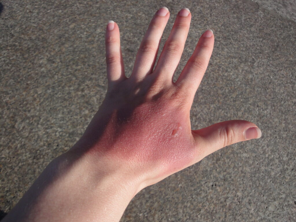
Hypertrophic scars occur when the body overproduces collagen, which causes the scar to be raised above the surrounding skin. Hypertrophic scars take the form of a red raised lump on the skin for lighter pigmented skin and the form of dark brown for darker pigmented skin. They usually occur within 4 to 8 weeks following wound infection or wound closure with excess tension and/or other traumatic skin injuries.
Keloid
Keloid scars are a more serious form of excessive scarring, because they can grow indefinitely into large, tumorous (although benign) neoplasms.
Hypertrophic scars are often distinguished from keloid scars by their lack of growth outside the original wound area, but this commonly taught distinction can lead to confusion.
Keloid scars can occur on anyone, but they are most common in dark-skinned people. They can be caused by surgery, cuts, accident, acne or, sometimes, body piercings. In some people, keloid scars form spontaneously. Although they can be a cosmetic problem, keloid scars are only inert masses of collagen and therefore completely harmless and not cancerous. However, they can be itchy or painful in some individuals. They tend to be most common on the shoulders and chest. Hypertrophic scars and keloids tend to be more common in wounds closed by secondary intention. Surgical removal of keloid is risky and may exacerbate the condition and worsening of the keloid.
Atrophic

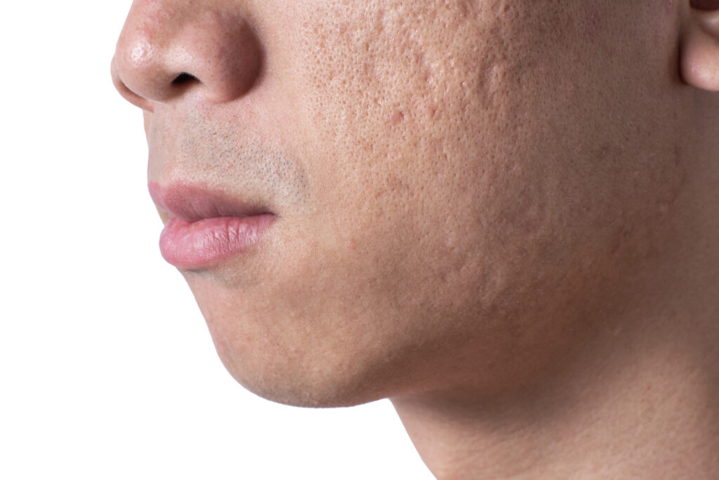
An atrophic scar takes the form of a sunken recess in the skin, which has a pitted appearance. These are caused when underlying structures supporting the skin, such as fat or muscle, are lost. This type of scarring is often associated with acne, chickenpox, other diseases (especially Staphylococcus infection), surgery, certain insect and spider bites, or accidents. It can also be caused by a genetic connective tissue disorder, such as Ehlers–Danlos syndrome.[11]
Stretch marks
Stretch marks (technically called striae) are also a form of scarring. These are caused when the skin is stretched rapidly (for instance during pregnancy, significant weight gain, or adolescent growth spurts), or when skin is put under tension during the healing process (usually near joints). This type of scar usually improves in appearance after a few years.
Elevated corticosteroid levels are implicated in striae development.
Umbilical
Humans and other placental mammals have an umbilical scar (commonly referred to as a navel) which starts to heal when the umbilical cord is cut after birth. Egg-laying animals have an umbilical scar which, depending on the species, may remain visible for life or disappear within a few days after birth.
Pathophysiology

A scar is the product of the body’s repair mechanism after tissue injury. If a wound heals quickly within two weeks with new formation of skin, minimal collagen will be deposited and no scar will form. When the extracellular matrix senses elevated mechanical stress loading, tissue will scar, and scars can be limited by stress shielding wounds. Small full thickness wounds under 2mm reepithelize fast and heal scar free. Deep second-degree burns heal with scarring and hair loss. Sweat glands do not form in scar tissue, which impairs the reguation of body temperature. Elastic fibers are generally not detected in scar tissue younger than 3 months old. In scars, rete pegs are lost; through a lack of rete pegs, scars tend to shear easier than normal tissue.
The endometrium, the inner lining of the uterus, is the only adult tissue to undergo rapid cyclic shedding and regeneration without scarring, shedding and restoring roughly inside a 7-day window on a monthly basis. All other adult tissues, upon rapid shedding or injury, can scar.
Prolonged inflammation, as well as the fibroblast proliferation,[25] can occur. Redness that often follows an injury to the skin is not a scar and is generally not permanent (see wound healing). The time it takes for this redness to dissipate may, however, range from a few days to, in some serious and rare cases, a few years.[26][citation needed]
Scars form differently based on the location of the injury on the body and the age of the person who was injured.[citation needed]
The worse the initial damage is, the worse the scar will generally be.[citation needed]
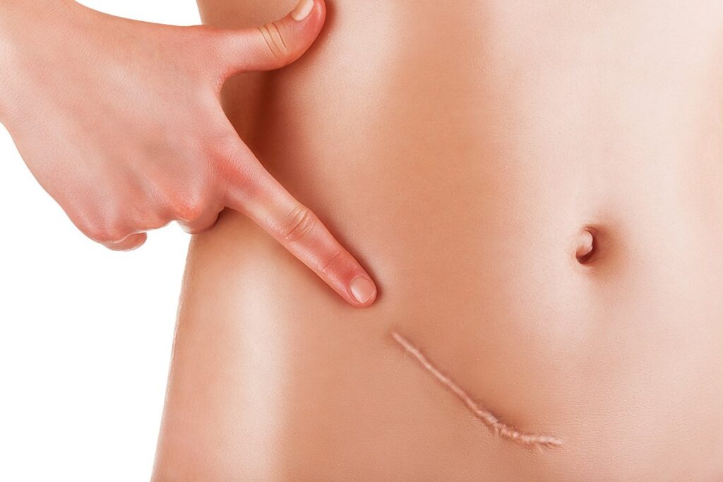
Skin scars occur when the dermis (the deep, thick layer of skin) is damaged. Most skin scars are flat and leave a trace of the original injury that caused them.
Wounds allowed to heal secondarily tend to scar worse than wounds from primary closure.
Collagen synthesis
An injury does not become a scar until the wound has completely healed; this can take many months, or years in the worst pathological cases, such as keloids. To begin to patch the damage, a clot is created; this clot is the beginning process that results in a provisional matrix. In the process, the first layer is a provisional matrix and is not a scar. Over time, the wounded body tissue overexpresses collagen inside the provisional matrix to create a collagen matrix. This collagen overexpression continues and crosslinks the fiber arrangement inside the collagen matrix, making the collagen dense. This densely packed collagen, morphing into an inelastic whitish collagen[25] scar wall, blocks off cell communication and regeneration; as a result, the new tissue generated will have a different texture and quality than the surrounding unwounded tissue. This prolonged collagen-producing process results in a fortuna scar.
Fibroblasts
The scarring is created by fibroblast proliferation,[25] a process that begins with a reaction to the clot.[27] To mend the damage, fibroblasts slowly form the collagen scar. The fibroblast proliferation is circular[27] and cyclically, the fibroblast proliferation lays down thick, whitish collagen[25] inside the provisional and collagen matrix, resulting in the abundant production of packed collagen on the fibers giving scars their uneven texture. Over time, the fibroblasts continue to crawl around the matrix, adjusting more fibers and, in the process, the scarring settles and becomes stiff.[27] This fibroblast proliferation also contracts the tissue.[27] In unwounded tissue, these fibers are not overexpressed with thick collagen and do not contract.
EPF and ENF fibroblasts have been genetically traced with the Engrailed-1 genetic marker.[28] EPFs are the primary contributors to all fibrotic outcomes after wounding.[28] ENFs do not contribute to fibrotic outcomes.
Myofibroblast
Mammalian wounds that involve the dermis of the skin heal by repair, not regeneration (except in 1st trimester inter-uterine wounds and in the regeneration of deer antlers). Full-thickness wounds heal by a combination of wound contracture and edge re-epitheliasation. Partial thickness wounds heal by edge re-epithelialisation and epidermal migration from adnexal structures (hair follicles, sweat glands and sebaceous glands). The site of keratinocyte stem cells remains unknown but stem cells are likely to reside in the basal layer of the epidermis and below the bulge area of hair follicles.
The fibroblast involved in scarring and contraction is the myofibroblast,[30] which is a specialized contractile fibroblast.[31] These cells express α-smooth muscle actin (α-SMA).[19] The myofibroblasts are absent in the first trimester in the embryonic stage where damage heals scar-free;[19] in small incisional or excision wounds less than 2 mm that also heal without scarring;[19] and in adult unwounded tissues where the fibroblast in itself is arrested; however, the myofibroblast is found in massive numbers in adult wound healing which heals with a scar.[31]
The myofibroblasts make up a high proportion of the fibroblasts proliferating in the postembryonic wound at the onset of healing. In the rat model, for instance, myofibroblasts can constitute up to 70% of the fibroblasts,[30] and is responsible for fibrosis on tissue.[32] Generally, the myofibroblasts disappear from the wound within 30 days,[33] but can remain in pathological cases in hypertrophy, such as keloids. Myofibroblasts have plasticity and in mice can be transformed into fat cells, instead of scar tissue, via the regeneration of hair follicles.
Mechanical stress
Wounds under 2mm generally do not scar but larger wounds generally do scar. In 2011 it was found that mechanical stress can stimulate scarring[18] and that stress shielding can reduce scarring in wounds. In 2021 it was found that using chemicals to manipulate fibroblasts to not sense mechanical stress brought scar-free healing. The scar-free healing also occurred when mechanical stress was placed onto a wound.
Treatment
Early and effective treatment of acne scarring can prevent severe acne and the scarring that often follows. In 2004, no prescription drugs for the treatment or prevention of scars were available.
Chemical peels
Chemical peels are chemicals which destroy the epidermis in a controlled manner, leading to exfoliation and the alleviation of certain skin conditions, including superficial acne scars.[40] Various chemicals can be used depending upon the depth of the peel, and caution should be used, particularly for dark-skinned individuals and those individuals susceptible to keloid formation or with active infections.[41]
Filler injections
Filler injections of collagen can be used to raise atrophic scars to the level of surrounding skin.[42] Risks vary based upon the filler used, and can include further disfigurement and allergic reaction.
Laser treatment
Nonablative lasers, such as the 585 nm pulsed dye laser, 1064 nm and 1320 nm Nd:YAG, or the 1540 nm Er:Glass are used as laser therapy for hypertrophic scars and keloids.[44] There is tentative evidence for burn scars that they improve the appearance.
Ablative lasers such as the carbon dioxide laser (CO2) or Er:YAG offer the best results for atrophic and acne scars.[47] Like dermabrasion, ablative lasers work by removing the epidermis.[48][49] Healing times for ablative therapy are much longer and the risk profile is greater compared to nonablative therapy; however, nonablative therapy offers only minor improvements in cosmetic appearance of atrophic and acne scars.[44] Combination laser therapy and microneedling may offer superior results to single modality treatment. The biggest recent advance in scar management is the use of fractionated CO2 laser and immediate application of topical steroid Triamcinolone.
Radiotherapy
Low-dose, superficial radiotherapy is sometimes used to prevent recurrence of severe keloid and hypertrophic scarring. It is thought to be effective despite a lack of clinical trials, but only used in extreme cases due to the perceived risk of long-term side effects.[50]
Dressings and topical silicone
Silicone scar treatments are commonly used in preventing scar formation and improving existing scar appearance.[51] A meta-study by the Cochrane collaboration found weak evidence that silicone gel sheeting helps prevent scarring.[52] However, the studies examining it were of poor quality and susceptible to bias.[52]
Pressure dressings are commonly used in managing burn and hypertrophic scars, although supporting evidence is lacking.[53] Care providers commonly report improvements, however, and pressure therapy has been effective in treating ear keloids.[53] The general acceptance of the treatment as effective may prevent it from being further studied in clinical trials.[53]
Verapamil-containing silicone gel
Verapamil, a type of calcium channel blocker, is considered a candidate drug for the treatment of hypertrophic scars. A study conducted by the Catholic University of Korea concluded that verapamil-releasing silicone gel is effective and is a superior alternative to the conventional silicone gel where decreased median SEI, fibroblast count, and collagen density in all verapamil-added treatment groups were observed.[54]: 647–656 Gross morphologic features suggested that the combination of verapamil and silicone improves the overall quality of hypertrophic scars by reducing scar height and redness. This was verified with quantifiable histomorphometric parameters; however, oral verapamil is not a good choice because of its effect of lowering blood pressure. Intralesional injection of verapamil is also suboptimal because of the required frequency for injections. Topical silicone gel combined with verapamil does not lead to systemic hypotension, is convenient to apply, and shows enhanced results.[54]: 647–656
Steroids
A long-term course of corticosteroid injections into the scar may help flatten and soften the appearance of keloid or hypertrophic scars.[55]
Topical steroids are ineffective.[56] However, clobetasol propionate can be used as an alternative treatment for keloid scars.[57]
Topical steroid applied immediately after fractionated CO2 laser treatment is however very effective (and more efficacious than laser treatment alone) and has shown benefit in numerous clinical studies.
Surgery

Scar revision is a process of cutting the scar tissue out. After the excision, the new wound is usually closed up to heal by primary intention, instead of secondary intention. Deeper cuts need a multilayered closure to heal optimally, otherwise depressed or dented scars can result.
Surgical excision of hypertrophic or keloid scars is often associated to other methods, such as pressotherapy or silicone gel sheeting. Lone excision of keloid scars, however, shows a recurrence rate close to 45%. A clinical study is currently ongoing to assess the benefits of a treatment combining surgery and laser-assisted healing in hypertrophic or keloid scars.
Subcision is a process used to treat deep rolling scars left behind by acne or other skin diseases. It is also used to lessen the appearance of severe glabella lines, though its effectiveness in this application is debatable. Essentially the process involves separating the skin tissue in the affected area from the deeper scar tissue. This allows the blood to pool under the affected area, eventually causing the deep rolling scar to level off with the rest of the skin area. Once the skin has leveled, treatments such as laser resurfacing, microdermabrasion or chemical peels can be used to smooth out the scarred tissue.
Vitamins
Research shows the use of vitamin E and onion extract (sold as Mederma) as treatments for scars is ineffective. Vitamin E causes contact dermatitis in up to 33% of users and in some cases it may worsen scar appearance and could cause minor skin irritations, but Vitamin C and some of its esters fade the dark pigment associated with some scars.


What is a scar?

There is a genetic predisposition in some people to produce thicker, itchy, enlarging scars called keloids.
Scarring is the process by which wounds are repaired. Damage to the deeper layer of the skin, the dermis, is required to produce a scar. Damage to only the epidermis, the most superficial layer of skin, will not always produce a scar.
Scars produce a structural change in the deeper layers of the skin which is perceived as an alteration in the architecture of the normal surface features. It is not just a change in skin color.
Fetal tissues and mucosal tissues can heal without producing a scar. Understanding how and why this is possible could lead to better surgical scar outcomes.
What are the different types of scars?
There is only one type of scar. The appearance of a scar depends on the nature of the wound that produced the damage, the anatomical location of the wound, and a variety of genetic factors that are different for each individual.
A defective healing process can result in a keloid, an unsightly, itchy, thick, red, knobby bump that often continues to enlarge over time. Keloids often are larger than the margins of the original wound.
What causes a scar?
The normal healing process in human tissue results in a scar. Scars occur when tissues have been significantly damaged and repaired. It can occur after physical trauma or as part of a disease process. Poorly controlled wound healing can result in thick, unsightly scars that cause symptoms. Therefore, when wounds are produced surgically, physicians utilize techniques to minimize scarring.
What are symptoms and signs of a scar?
Scars occur at the site of tissue damage and appear as firm red to purple fibrous tissue that over time usually becomes flatter and lighter in color.
How do healthcare professionals diagnose scars?
Scars are almost always diagnosed by visual inspection. There are several rare situations when it may be necessary to examine scar tissue under a microscope to confirm its true identity. This would require a biopsy of the skin and may require the injection of a local anesthetic.
Sometimes other skin conditions can form in a scar and require a biopsy to be diagnosed.
What is the treatment for a scar?
Since scars are part of the normal healing process, ordinary scars are not treated. Only when superficial scars become cosmetically undesirable do they require treatment. This would include scars in those who are predisposed to develop keloids, as well as scars in anatomical regions known to produce thick scars and scars that produce a significant, unpleasant distortion of adjacent anatomical structures.
Thick scars and keloids often flatten out after injections of steroids directly into the fibrous scar tissue. They can respond to the chronic application of pressure and the application of sheets of silicone rubber. Thick scars can be flattened by dermabrasion, which utilizes abrasive devices to sand down elevated scars.
Certain types of depressed scars can be elevated by the injection of a cosmetic skin filler. Certain types of facial scarring respond well to forms of laser treatments. Occasionally, surgical revision of scars can result in a different scar that is much more cosmetically desirable.
Since it takes about a year for scars to mature, it is frequently prudent to wait before starting any invasive surgical revisions.
Are there any home remedies to reduce scarring?
Good wound care is important in preventing excessive scarring as well as speeding up the healing process. Preventing infection can help prevent unnecessary inflammation which can increase the size of wounds resulting in larger, unsightly scars. It is important to remove crusts (scabs) from wounds gently with a washcloth and soap and water at least twice a day and to keep the wound moist by keeping it covered with petroleum jelly or antibiotic ointment, such as Neosporin.
Assuming the wound is healing normally, it would not be unreasonable to cover the wound site after it is covered by skin (epithelialized) with silicone rubber 24 hours a day for a month or so. There is medical evidence that this can diminish the thickness of scars. Proprietary products of this type can be purchased without a prescription. There is an over-the-counter product (Mederma) that may improve the appearance of scars in the short term (first one to two months) but conclusive evidence of efficacy is lacking. This product relies on an extract of onions.
The judicious use of cosmetic makeup can effectively obscure many scars.
What is the prognosis of a scar?
Scars generally improve in appearance over the first year. So considerations for invasive treatments need to be prudently considered before that time. On the other hand, scarring usually involves tissue contraction, so that it is unlikely that scars that pull or twist other anatomical structures, producing unpleasant results, will improve. These should be treated sooner than later.
Is it possible to prevent scarring?
Scarring is an integral part of the healing process. Assuming the wound does not become infected, physicians plan excisions to minimize the cosmetic defects produced by scars. This can be accomplished by orienting the wound in such a way that it will not perturb other structures so that the scar can be camouflaged by hiding in wrinkle lines or near other anatomical structures. It is also important to minimize the tension necessary to close the wound surgically.
Does insurance coverage apply to scar treatments?
Most medical insurance does not cover cosmetic procedures. If scarring has produced a change that is deemed other than cosmetic, it is reasonable to expect coverage, for example, when scarring is the result of trauma. Occasionally, this question may be open to dispute so it can be helpful to have a physician’s office intercede with the carrier before performing the procedure.
Skin Problems: Rosacea, Acne, Shingles, Covid-19 Rashes
Skin Problems?

Is your skin itchy, oozing, or breaking out? Moles, psoriasis, hives, eczema, and recently associated Covid-19 coronavirus rashes are just a few of the more than 3,000 skin disorders known to dermatology. Changes in color or texture can result from inflammation, infection, or allergic reactions anywhere on the body. Some skin conditions can be minor, temporary, and easily treated — while others can be very serious, and even life-threatening. Read on to see signs and symptoms of the most common skin disorders and learn how to identify them.
Covid-19 (Coronavirus) Skin Rashes

Skin rashes have been associated with COVID-19 infection. Much like other viral diseases such as HIV and bacterial diseases like syphilis, COVID-19 rashes can take many different forms. One study from Spain identified five patterns of COVID-19 rash. The most common type was a “macropapular rash.” These rashes feature both small, flat discolorations (“macules”) and small, elevated lesions (“papule”). These rashes are associated with more severe COVID-19 infection, as 2% of those who got them in the Spain study reportedly died from the illness. Other rashes associated with COVID-19 include thickened lesions developing on the heels of the feet, lesions that resemble chickenpox, and rashes that resemble those seen with dengue fever.
Some dermatologists have reported cases of so-called “COVID toe” in both adults and children. These lesions may be reddish, elevated lesions that flatten after about a week. Some of the patients found their COVID toe rashes itchy, and others did not. Some found it painful when their toes were pressed, and others did not. More research is needed, as some of the rashes reported in COVID-19 patients resemble drug reactions. For safety reasons, researchers have been unable to determine if drug interactions are responsible in these cases, or whether the novel coronavirus itself causes these rashes.
Shingles (Herpes Zoster)

Shingles, also known as herpes zoster, is a skin disease caused by the return of a chickenpox infection from latently infected nerve cells in the spinal cord or brain. It begins as a painful sensation which is often mistaken for a musculoskeletal injury or even a heart attack. It is soon followed within one or two days by a red, blistering unilateral (one-sided) rash distributed to the skin supplied by a sensory nerve (a dermatome). Zoster tends to occur most often in the elderly and can be largely prevented or made less severe with a vaccination. Treatment with antiviral drugs within 48 hours of the onset of the eruption may limit the development of a persistent, severe pain (neuralgia) at the site of the eruption.
Hives (Urticaria)

Hives, also known as urticaria, is one of the most common allergic skin conditions. It most often occurs due to antibodies in the bloodstream that recognize foreign substances. This eruption appears suddenly anywhere on the body as elevated blanched bumps surrounded by an intensely itchy red rash. There may be many lesions, but each one only exists for eight to 12 hours. As older ones resolve, newer ones may develop. Most of the time, urticaria resolves spontaneously within eight weeks and is treated with oral antihistamines for symptomatic relief.
Psoriasis

Psoriasis is a chronic, inflammatory genetic condition in which patients develop scaly red bumps that coalesce into plaques. Symptoms of psoriasis typically occur but are not limited to the scalp, elbows, and knees.
Psoriasis is not curable; flare-ups come and go by themselves. There are a variety of treatments depending on the severity and extent of involvement, which vary from topical creams and ultraviolet light exposure to oral drugs and injectable medications. Patients with psoriasis more commonly develop cardiovascular disease and diabetes, which may be attributable to system-wide inflammation.
Eczema (Dermatitis)

Eczema (sometimes called “dermatitis”) is a genetic condition associated with itchy, dry skin. It usually develops in early childhood with symptoms of a chronically itchy, weeping, oozing sores. Eczema tends to be found on arm creases opposite the elbow and on leg creases opposite the knee.
Many eczema patients also have inhalant allergies such as asthma and hay fever. Eczema improves with age. Treatment involves applying emollients to wet skin and using topical steroids.
Types of Eczema
There are many types of eczema, and many types include the word “dermatitis” (in dermatology, dermatitis is another word for eczema). For instance, eczema types include stasis dermatitis and dyshidrotic eczema. A dermatologist can help you understand what type you have. Two of the most common types are:
- Atopic dermatitis
- Contact dermatitis
Rosacea

Rosacea is a chronic inflammatory condition of the face that is characterized by redness, dilated blood vessels, papules, pustules, and occasionally by the overgrowth of nasal connective tissue (rhinophyma). It superficially resembles teenaged acne, but it occurs in adults. Persistent facial flushing is an early sign of the skin’s uncontrolled sensitivity to certain naturally produced inflammatory chemicals. Treatment of rosacea involves topical and oral drugs.
Cold Sores (Fever Blisters)

Herpes labialis (cold sore) is caused by the herpes simplex virus. Cold sores commonly appear on the edge of the lip. This virus exists in a dormant state in the spinal cord nerve cells, and after certain environmental triggers like a sunburn or a cold, the virus is induced to travel along a peripheral nerve to the same skin site over and over again. The eruption is self-limited to about seven to 10 days so that treatment is unnecessary unless the eruption becomes too frequent.
Plant Rashes

In allergic individuals, the development of a linear blistering eruption occurs within 24-48 hours of exposure to a member of the poison ivy or poison oak family of plants. Since the plant contains highly allergenic chemicals, most people will become allergic after a single priming exposure. The eruption will resolve within three weeks but will occur again the next time the skin comes in contact with the plant.
Treating Plant Rashes

The repeated application of cool wet compresses to the blisters followed by evaporation of the water can be soothing and speed healing. Treatment with steroids creams or even oral steroids may be required in severe cases. Once a person is allergic, this is permanent; it is important to avoid this plant family assiduously so this very unpleasant allergic reaction will not recur. Many of those allergic to poison ivy or poison oak (Toxicodendron) are also sensitive to mango skin and cashew nut oil.
Razor Bumps

This eruption occurs in areas of the skin in which hairs have been recently cut or extracted. This is commonly present in the beard area of individuals with very tightly coiled hair. When the hair is cut off or plucked out below the level of the follicular pore, it tends to curl into the side of the follicle and cause an inflammatory bump. Not shaving closely is very important in preventing this skin condition.
Skin Tags

Skin tags are small, fleshy, fibrovascular, pedunculated (on a stalk) growths that are often are found on the neck and armpits. They are generally asymptomatic unless they become irritated by frictional forces or their blood supply becomes compromised. They are very common and need not be removed or destroyed unless they become irritated.
Acne

Acne vulgaris is usually a noninfectious eruption of papules and pustules (pus-filled blisters) on the face and occasionally on the chest and back. Acne occurs in all teenagers as they progress through puberty. Symptoms like comedones (blackheads) and inflammatory papules and pustules all appear simultaneously.
Despite rumors to the contrary, acne is not caused by dirty skin. Instead, it is mediated by hormones that begin to circulate during puberty and excess sebum or oil production. The condition generally resolves around the age of 20-30 but may produce scarring if severe and left untreated.
Athlete’s Foot

One of the most commonplace skin conditions is athlete’s foot. And one of the most common causes of athlete’s foot is an infection of the dead superficial layer of the skin called the stratum corneum by a fungal mold (tinea pedis) called a dermatophyte.
If inflammatory, this condition may cause fluid-filled blisters that are quite itchy. Noninflammatory tinea pedis produces scaly, dry skin. Often it is only mildly irritating. Tinea pedis is probably frequently contracted by walking barefoot in locker rooms. Topical antifungal creams are available over the counter and can be helpful in treating this skin infection. More powerful medications can be prescribed by a dermatologist.
Moles

Although the term mole may cover a variety of different sorts of skin growths, most often it refers to a localized accumulation of pigment-producing cells called melanocytes. These are generally uniform in color and round in shape. In dermatology, moles are sometimes known as benign neoplasms.
Melanocytic nevi (moles) range in color from beige to black, they’re about ½ an inch in diameter, and are often located on sun-exposed skin. Poorly pigmented individuals may have an average of 35 of these growths by the time they are 35 years old. These are benign lesions but can be confused with various pigmented skin cancers. Pigmented lesions that itch, bleed, or grow could be cause for concern.
Age or Liver Spots

Liver spots (also called age spots) are a common skin condition that typically appears on the face and forearms of older individuals. Although these flat brown spots cause no symptoms, patients detest them because of their unsightly appearance. They can be treated in a variety of ways, but treatment is not medically necessary.
Pityriasis Rosea

This rash usually begins in a young adult as a single scay bump or patch and then extends to cover much of the torso with many scaly spots that are elliptical in shape. They are associated with modest itching which only occasionally requires treatment. The condition usually lasts about 6-8 weeks in total.
Melasma

Melasma is another commonly experienced skin condition. The main symptoms are brown patches of skin. These patches are typically found on your face.
This condition occurs most commonly in women of childbearing age and is often associated with pregnancy or the ingestion of oral contraceptive medication. This flat brownish pigmentation occurs on the forehead, cheeks, and in the mustache area of the upper lip. It often persists after pregnancy or after birth control has ceased. Sunlight will make it darker. Successful treatment is not easy, and strict sun protection is a necessity.
Warts

The development of small keratotic tumors of the skin is caused by one of about 200 members of the human papillomavirus group. They often spontaneously go away, but particularly stubborn warts may require medical intervention. The proliferation of various treatments reflects the fact that successful resolution mostly depends upon the patient’s immune response. There are a variety of treatments available without a prescription that ought to be tried prior to seeing a physician.
Seborrheic Keratoses

This is the single most common benign bump present on people as they age. (Benign means it does not indicate skin cancer). Lesions may be present anywhere on the body and generally do not produce symptoms. They appear as black, brown, or yellow bumpy lesions which give the appearance of having been “glued” onto the skin. They are of no medical significance aside from the fact that they are occasionally confused with pigmented skin cancers.
Seborrheic Dermatitis

Seborrheic dermatitis is the single most common rash of adults. When it occurs in infancy, it is commonly called cradle cap. The adult disease tends to favor the scalp, skin behind the ears, forehead, brows, nasolabial folds of the face, mid-chest area, and the mid-back, producing an itchy, red scaling dermatitis. The scaling in the scalp can be conspicuous, producing impressive dandruff. The cause of this condition is unclear, but it responds well to topical steroids and to topical antifungal creams. Medicated shampoos containing tar, selenium sulfide, and zinc pyrithione are often effective. This condition commonly improves spontaneously but will ultimately recur. There is no cure so treatment must continue indefinitely.


